Showing posts with label Nursing Care Plan. Show all posts

Wednesday, September 22, 2010

Nursing Care Plan for Dengue Hemorrhagic Fever (DHF)

Nursing Care Plan for Dengue Hemorrhagic Fever (DHF)

Dengue Hemorrhagic Fever

Dengue hemorrhagic fever is a severe, potentially deadly infection spread by certain species of mosquitoes (Aedes aegypti).


Causes

Four different dengue viruses are known to cause dengue hemorrhagic fever. Dengue hemorrhagic fever occurs when a person catches a different type dengue virus after being infected by another one sometime before. Prior immunityimmunity to a different dengue virus type plays an important role in this severe disease.

Worldwide, more than 100 million cases of dengue fever occur every year. A small number of these develop into dengue hemorrhagic fever. Most infections in the United States are brought in from other countries. It is possible for a traveler who has returned to the United States to pass the infection to someone who has not traveled.

Risk factors for dengue hemorrhagic fever include having antibodies to dengue virus from prior infection and being younger than 12, female, or Caucasian.


Symptoms

Early symptoms of dengue hemorrhagic fever are similar to those of dengue fever, but after several days the patient becomes irritable, restlessrestless, and sweaty. These symptoms are followed by a shockshock -like state.

Bleeding may appear as tiny spots of blood on the skin (petechiaepetechiae) and larger patches of blood under the skin (ecchymosesecchymoses). Minor injuries may cause bleeding.

Shock may cause death. If the patient survives, recovery begins after a one-day crisis period.

Early symptoms include:

* Decreased appetite
* Fever
* Headache
* Joint aches
* MalaiseMalaise
* Muscle aches
* Vomiting

AcuteAcute phase symptoms include:

* Restlessness followed by:
o Ecchymosis
o Generalized rash
o Petechiae
o Worsening of earlier symptoms
* Shock-like state
o Cold, clammy extremities
o Sweatiness (diaphoretic)


Nursing Care Plan for Dengue Hemorrhagic Fever (DHF)


Assessment
Assessment is the initial phase of the nurse to obtain the required data before performing nursing care. assessment in patients with "DHF" can be done with the interview technique, measurement, and physical examination. As for step-stages include:
* Identify potential sources and are available to meet patient needs.
* Assess the patient's medical history.
* Assess the increase in body temperature, signs of hemorrhage, nausea, vomiting, no appetite, heartburn, sore muscles and joints, signs of shock (rapid and weak pulse, hypotension, cold and moist skin, especially on the extremities, cyanosis, agitation, decreased consciousness).


Nursing Diagnosis and Intervention

1. Disorders of body fluid volume is less than body requirements related to increased capillary permeability, bleeding, vomiting and fever.

Objective: Disorders of body fluid volume can be solved
Result Criteria :
* Volume of body fluids back to normal

Intervention :
* Assess the patient's general condition and the condition
* Observation of vital signs (Temperature, Pulse)
Observation * signs of dehydration
* Observations drip infusion, and the location of the insertion of intravenous needles
* Balance fluid (the fluid input and output)
* Give the patient and family encourage patients to drink plenty
* Instruct the patient's family to change his clothes soaked in patients
sweat.

Nursing Care Plan for Dengue Hemorrhagic Fever (DHF)

Nursing Care Plan for Dengue Hemorrhagic Fever (DHF)

Dengue Hemorrhagic Fever

Dengue hemorrhagic fever is a severe, potentially deadly infection spread by certain species of mosquitoes (Aedes aegypti).


Causes

Four different dengue viruses are known to cause dengue hemorrhagic fever. Dengue hemorrhagic fever occurs when a person catches a different type dengue virus after being infected by another one sometime before. Prior immunityimmunity to a different dengue virus type plays an important role in this severe disease.

Worldwide, more than 100 million cases of dengue fever occur every year. A small number of these develop into dengue hemorrhagic fever. Most infections in the United States are brought in from other countries. It is possible for a traveler who has returned to the United States to pass the infection to someone who has not traveled.

Risk factors for dengue hemorrhagic fever include having antibodies to dengue virus from prior infection and being younger than 12, female, or Caucasian.


Symptoms

Early symptoms of dengue hemorrhagic fever are similar to those of dengue fever, but after several days the patient becomes irritable, restlessrestless, and sweaty. These symptoms are followed by a shockshock -like state.

Bleeding may appear as tiny spots of blood on the skin (petechiaepetechiae) and larger patches of blood under the skin (ecchymosesecchymoses). Minor injuries may cause bleeding.

Shock may cause death. If the patient survives, recovery begins after a one-day crisis period.

Early symptoms include:

* Decreased appetite
* Fever
* Headache
* Joint aches
* MalaiseMalaise
* Muscle aches
* Vomiting

AcuteAcute phase symptoms include:

* Restlessness followed by:
o Ecchymosis
o Generalized rash
o Petechiae
o Worsening of earlier symptoms
* Shock-like state
o Cold, clammy extremities
o Sweatiness (diaphoretic)


Nursing Care Plan for Dengue Hemorrhagic Fever (DHF)


Assessment
Assessment is the initial phase of the nurse to obtain the required data before performing nursing care. assessment in patients with "DHF" can be done with the interview technique, measurement, and physical examination. As for step-stages include:
* Identify potential sources and are available to meet patient needs.
* Assess the patient's medical history.
* Assess the increase in body temperature, signs of hemorrhage, nausea, vomiting, no appetite, heartburn, sore muscles and joints, signs of shock (rapid and weak pulse, hypotension, cold and moist skin, especially on the extremities, cyanosis, agitation, decreased consciousness).


Nursing Diagnosis and Intervention

1. Disorders of body fluid volume is less than body requirements related to increased capillary permeability, bleeding, vomiting and fever.

Objective: Disorders of body fluid volume can be solved
Result Criteria :
* Volume of body fluids back to normal

Intervention :
* Assess the patient's general condition and the condition
* Observation of vital signs (Temperature, Pulse)
Observation * signs of dehydration
* Observations drip infusion, and the location of the insertion of intravenous needles
* Balance fluid (the fluid input and output)
* Give the patient and family encourage patients to drink plenty
* Instruct the patient's family to change his clothes soaked in patients
sweat.

Sunday, August 8, 2010

Nursing Care Plan for Schizophrenia

NCP - Nursing Care Plan for Schizophrenia

Schizophrenia


Basic Concept of Schizophrenia

Definition

Schizophrenia is a syndrome with various descriptions of the cause (many not yet known) and history (not necessarily chronic or deteriorating) wide, and a number of consequences which depend on consideration of the influence of genetic, physical and socio-cultural (Rusdi Maslim, 1997; 46).

Causes
Offspring
It has been proven by research that the morbidity rate for the half-brother from 0.9 to 1.8%, 7-15% for siblings, for children with one parent who suffered from Schizophrenia 40-68%, 2-15% twins two eggs and one egg twins 61-86% (Maramis, 1998; 215).

Endocrine
This theory is put forward in connection with the frequent occurrence of Schizophrenia at the time of puberty, pregnancy or puerperium time and time climacterium., But this theory can not be proven.

Metabolism
This theory is based for Schizophrenia patients appear pale, unhealthy, a little tip extremity cyanosis, decreased appetite and weight declined and catatonic stupor in patients with decreased oxygen consumption. This hypothesis is still in the proof with the hallucinogenic drug administration.

Central nervous system
Schizophrenia Causes CNS disorders is directed at the diensefalon or brain cortex, but the pathological abnormalities found may be due to postmortem changes or is artefakt in time to make preparations.


Assessment

Assessment is the beginning and the main basis of the assessment phase of nursing process consists of collecting data and formulation of clients' needs or problems.

The data collected consist of biological, psychological, social and spiritual. Grouping of data on mental health pengakajian can also be the predisposing factor, precipitation factor, the assessment of stressors, coping resources and coping abilities possessed clients (stuart and Sunden, 1998). Other studies focus on the way 5 (five) dimensions: physical, emotional, intellectual, social and spiritual. The contents of the study include :
  1. Client identity
  2. The main complaint / reason for entry
  3. Predisposing factors
  4. Dimensional physical / biological
  5. Psychosocial dimensions
  6. Mental status
  7. Preparation needs to go home
  8. Coping mechanism
  9. Psychosocial and environmental problems
  10. Medical Aspects
The data obtained through direct observation or examination referred to objective data, the data is delivered in a talk to clients and families through the interview treatment called subjective data.


Nursing Diagnosis

Risk injuring themselves and or others / environment related to changes in sensory perception / hallucinations

General Objectives :
Clients do not mencideri themselves and or others / environment.

Specific objectives :
  1. Clients can be a trusting relationship :
    Nursing Intervention :
    • Construct a trusting relationship
      • Regards therapeutic
      • Introducing yourself
      • Explain the purpose of interaction
      • Create a calm environment
      • Create a contract that clearly at every meeting (topics, time and place to talk).
    • Give clients the opportunity to express his feelings.
    • Listen to the client expression of empathy.

  2. Clients can recognize hallucinations
    Nursing Intervention :
    • Make frequent contact and brief
      Rational: to reduce the client's contact with the hallucinations.
    • Obeservasi client behavior associated with hallucinations; talk and laugh without the stimulus, kesekitarnya looked as if there is someone to talk.
    • Help clients to know the hallucinations;
      • If the client answered no, proceed; what was said ?
      • Say that the nurse believes the client to hear.
      • Tell that other clients also have such clients.
      • Say that the treatment will help the client.
    • Discuss with the client about;
      • Situations that can cause / not cause hallucinations.
      • Time and frequency of occurrence of hallucinations (morning, afternoon, evening, night or when alone or when upset / sad).
    • Discuss with the client about what is felt when there hallucinations (angry / scared / sad / happy) and the opportunity to express feelings.

  3. Clients can control the hallucinations
    Nursing Intervention :
    • Identification with the client ways / actions taken when there hallucinations (sleep / angry / busy myself)
    • Discuss the benefits of the ways in which clients, if useful give a compliment.
    • Discussion of new ways to cut / control the occurrence of hallucinations :
      • Say "I do not want with you" (the hallucinations).
      • Meet with other people (nurses / friends / family members to talk to say hallucinations.
      • Create a schedule of daily activities - the day that hallucinations do not have time to appear.
      • Ask other people (nurses / friends of family members) say hello if looks speak for herself.
    • Help clients decide how to select and train / control the hallucinations gradually.
    • Give the opportunity to perform the way they are trained, evaluated the results and praise when successful.
    • Encourage clients to participate in group activity therapy (stimulation of the realization and perception of orientation).
NCP - Nursing Care Plan for Schizophrenia

Nursing Care Plan for Schizophrenia

NCP - Nursing Care Plan for Schizophrenia

Schizophrenia


Basic Concept of Schizophrenia

Definition

Schizophrenia is a syndrome with various descriptions of the cause (many not yet known) and history (not necessarily chronic or deteriorating) wide, and a number of consequences which depend on consideration of the influence of genetic, physical and socio-cultural (Rusdi Maslim, 1997; 46).

Causes
Offspring
It has been proven by research that the morbidity rate for the half-brother from 0.9 to 1.8%, 7-15% for siblings, for children with one parent who suffered from Schizophrenia 40-68%, 2-15% twins two eggs and one egg twins 61-86% (Maramis, 1998; 215).

Endocrine
This theory is put forward in connection with the frequent occurrence of Schizophrenia at the time of puberty, pregnancy or puerperium time and time climacterium., But this theory can not be proven.

Metabolism
This theory is based for Schizophrenia patients appear pale, unhealthy, a little tip extremity cyanosis, decreased appetite and weight declined and catatonic stupor in patients with decreased oxygen consumption. This hypothesis is still in the proof with the hallucinogenic drug administration.

Central nervous system
Schizophrenia Causes CNS disorders is directed at the diensefalon or brain cortex, but the pathological abnormalities found may be due to postmortem changes or is artefakt in time to make preparations.


Assessment

Assessment is the beginning and the main basis of the assessment phase of nursing process consists of collecting data and formulation of clients' needs or problems.

The data collected consist of biological, psychological, social and spiritual. Grouping of data on mental health pengakajian can also be the predisposing factor, precipitation factor, the assessment of stressors, coping resources and coping abilities possessed clients (stuart and Sunden, 1998). Other studies focus on the way 5 (five) dimensions: physical, emotional, intellectual, social and spiritual. The contents of the study include :
  1. Client identity
  2. The main complaint / reason for entry
  3. Predisposing factors
  4. Dimensional physical / biological
  5. Psychosocial dimensions
  6. Mental status
  7. Preparation needs to go home
  8. Coping mechanism
  9. Psychosocial and environmental problems
  10. Medical Aspects
The data obtained through direct observation or examination referred to objective data, the data is delivered in a talk to clients and families through the interview treatment called subjective data.


Nursing Diagnosis

Risk injuring themselves and or others / environment related to changes in sensory perception / hallucinations

General Objectives :
Clients do not mencideri themselves and or others / environment.

Specific objectives :
  1. Clients can be a trusting relationship :
    Nursing Intervention :
    • Construct a trusting relationship
      • Regards therapeutic
      • Introducing yourself
      • Explain the purpose of interaction
      • Create a calm environment
      • Create a contract that clearly at every meeting (topics, time and place to talk).
    • Give clients the opportunity to express his feelings.
    • Listen to the client expression of empathy.

  2. Clients can recognize hallucinations
    Nursing Intervention :
    • Make frequent contact and brief
      Rational: to reduce the client's contact with the hallucinations.
    • Obeservasi client behavior associated with hallucinations; talk and laugh without the stimulus, kesekitarnya looked as if there is someone to talk.
    • Help clients to know the hallucinations;
      • If the client answered no, proceed; what was said ?
      • Say that the nurse believes the client to hear.
      • Tell that other clients also have such clients.
      • Say that the treatment will help the client.
    • Discuss with the client about;
      • Situations that can cause / not cause hallucinations.
      • Time and frequency of occurrence of hallucinations (morning, afternoon, evening, night or when alone or when upset / sad).
    • Discuss with the client about what is felt when there hallucinations (angry / scared / sad / happy) and the opportunity to express feelings.

  3. Clients can control the hallucinations
    Nursing Intervention :
    • Identification with the client ways / actions taken when there hallucinations (sleep / angry / busy myself)
    • Discuss the benefits of the ways in which clients, if useful give a compliment.
    • Discussion of new ways to cut / control the occurrence of hallucinations :
      • Say "I do not want with you" (the hallucinations).
      • Meet with other people (nurses / friends / family members to talk to say hallucinations.
      • Create a schedule of daily activities - the day that hallucinations do not have time to appear.
      • Ask other people (nurses / friends of family members) say hello if looks speak for herself.
    • Help clients decide how to select and train / control the hallucinations gradually.
    • Give the opportunity to perform the way they are trained, evaluated the results and praise when successful.
    • Encourage clients to participate in group activity therapy (stimulation of the realization and perception of orientation).
NCP - Nursing Care Plan for Schizophrenia

Thursday, August 5, 2010

NCP - Nursing Care Plan for Child with Hydrocephalus

NCP - Nursing Care Plan for Child with Hydrocephalus

NCP Hydrocephalus

Hydrocephalus

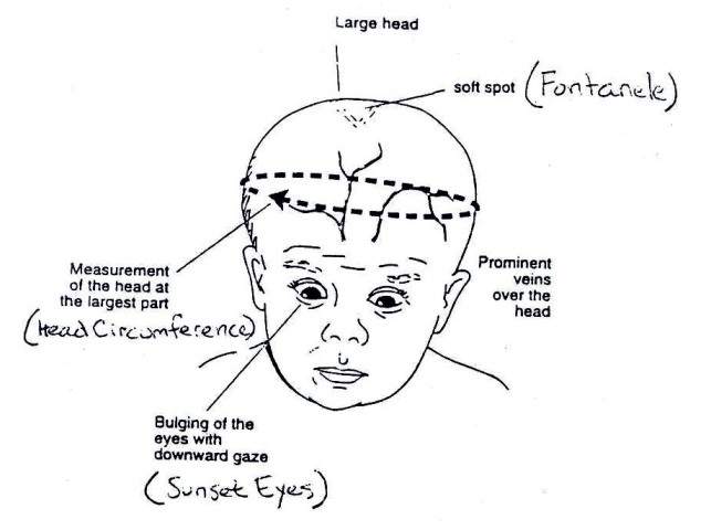
The term hydrocephalus is derived from the Greek words "hydro" meaning water and "cephalus" meaning head. As the name implies, it is a condition in which the primary characteristic is excessive accumulation of fluid in the brain. Although hydrocephalus was once known as "water on the brain," the "water" is actually cerebrospinal fluid (CSF) — a clear fluid that surrounds the brain and spinal cord. The excessive accumulation of CSF results in an abnormal widening of spaces in the brain called ventricles. This widening creates potentially harmful pressure on the tissues of the brain.

The ventricular system is made up of four ventricles connected by narrow passages.. Normally, CSF flows through the ventricles, exits into cisterns (closed spaces that serve as reservoirs) at the base of the brain, bathes the surfaces of the brain and spinal cord, and then reabsorbs into the bloodstream.

CSF has three important life-sustaining functions :
  1. to keep the brain tissue buoyant, acting as a cushion or "shock absorber";
  2. to act as the vehicle for delivering nutrients to the brain and removing waste; and
  3. to flow between the cranium and spine and compensate for changes in intracranial blood volume (the amount of blood within the brain).

The balance between production and absorption of CSF is critically important. Because CSF is made continuously, medical conditions that block its normal flow or absorption will result in an over-accumulation of CSF. The resulting pressure of the fluid against brain tissue is what causes hydrocephalus.
www.medicinenet.com

Clinical manifestations :
  • Enlarging head circumference
  • Irritable, lethargic, apathetic
  • Convergent strabismus
  • Increased Intra-cranial pressure, decreased consciousness
  • Fontanels: tense; bulging
  • Manifestation appropriate location in the brain lesions.

Assessment
  • Medical history: cerebral trauma, cerebral infections, etc.
  • Physical examination: a focus area of the head
  • Inspection psychomotor
  • Other clinical manifestations
  • Check diagnostic

Nursing Diagnosis
  • The risk of injury related to increased intra-cranial pressure
  • The risk of infection related to mechanical drainage installed
  • Changes in tissue perfusion related to interruption of blood flow
  • The risk of skin integrity related disorders with an emphasis on the area behind the head
  • Impaired family processes related to situational crises (children with physical disorders)

Nursing Intervention

The risk of injury associated with increased intra-cranial pressure
  • Observation of Increased intra cranial pressure
  • Perform neurological assessment
  • Position the patient with safe, elevate the head area
  • Avoid the use of sedation.

NCP - Nursing Care Plan for Child with Hydrocephalus

NCP - Nursing Care Plan for Child with Hydrocephalus

NCP Hydrocephalus

Hydrocephalus

The term hydrocephalus is derived from the Greek words "hydro" meaning water and "cephalus" meaning head. As the name implies, it is a condition in which the primary characteristic is excessive accumulation of fluid in the brain. Although hydrocephalus was once known as "water on the brain," the "water" is actually cerebrospinal fluid (CSF) — a clear fluid that surrounds the brain and spinal cord. The excessive accumulation of CSF results in an abnormal widening of spaces in the brain called ventricles. This widening creates potentially harmful pressure on the tissues of the brain.

The ventricular system is made up of four ventricles connected by narrow passages.. Normally, CSF flows through the ventricles, exits into cisterns (closed spaces that serve as reservoirs) at the base of the brain, bathes the surfaces of the brain and spinal cord, and then reabsorbs into the bloodstream.

CSF has three important life-sustaining functions :
  1. to keep the brain tissue buoyant, acting as a cushion or "shock absorber";
  2. to act as the vehicle for delivering nutrients to the brain and removing waste; and
  3. to flow between the cranium and spine and compensate for changes in intracranial blood volume (the amount of blood within the brain).

The balance between production and absorption of CSF is critically important. Because CSF is made continuously, medical conditions that block its normal flow or absorption will result in an over-accumulation of CSF. The resulting pressure of the fluid against brain tissue is what causes hydrocephalus.
www.medicinenet.com

Clinical manifestations :
  • Enlarging head circumference
  • Irritable, lethargic, apathetic
  • Convergent strabismus
  • Increased Intra-cranial pressure, decreased consciousness
  • Fontanels: tense; bulging
  • Manifestation appropriate location in the brain lesions.

Assessment
  • Medical history: cerebral trauma, cerebral infections, etc.
  • Physical examination: a focus area of the head
  • Inspection psychomotor
  • Other clinical manifestations
  • Check diagnostic

Nursing Diagnosis
  • The risk of injury related to increased intra-cranial pressure
  • The risk of infection related to mechanical drainage installed
  • Changes in tissue perfusion related to interruption of blood flow
  • The risk of skin integrity related disorders with an emphasis on the area behind the head
  • Impaired family processes related to situational crises (children with physical disorders)

Nursing Intervention

The risk of injury associated with increased intra-cranial pressure
  • Observation of Increased intra cranial pressure
  • Perform neurological assessment
  • Position the patient with safe, elevate the head area
  • Avoid the use of sedation.

Tuesday, June 8, 2010

Ncp - Nursing Care Plan Patient Heart Failure


NCP for Heart Failure

Assessment
  1. Left-sided heart failure ; Dyspnea, Crackles, Orthopnea, Paroxysmal noctural dyspnea, Tachypnea, Tachycardia, Gallop rhythm (third or S3 and fourth or S4 heart sound), Fatigue, Anxiety, Arrhythmias and Cough.
  2. Righ-sided heart failure ; Dependent edema, Weight gain, Fatique, Jugular vein distention, Tachycardia, Gallop rhythm (S3 or S4), Nausea, Anorexia, Hepatomegaly and Ascites.

Nursing Diagnoses
  • Excess fluid volume
  • Activity intolerance
  • Ineffective health maintenance

Planing and Goals of Nursing Care
  • The clients will understand how to cope with necessary lifestyle changes.
  • The client won't develop preventable complication
  • The client will will understand how to continue therapy at home.

Nursing Intervention For Heart Failure
  • Assess cardiovascular status, vital sign and hemodynamic variable to detect signs of reduced cardiac output.
  • Assess respiratory status to detect increasing fluid in the lungs and respiratory failure.
  • Keep the client in semi-fowler's position to increase chest expansion and improve ventilation.
  • Administer medication as prescribed, to enhance cardiac performance and reduce excess fluids.
  • Administer oxygen to enhance arterial oxygenation.
  • Measure and record intake and output, Intake greater than output may indicated fluid retention.
  • Monitor laboratory test result to detect electrolyte imbalances, renal failure, and impaired cardiac circulation.
  • Provide suctioning, if necessary assist with turning and encourage coughing and deep breathing to prevent pulmonary complication.
  • Restrict oral fluid to avoid worsening the client's condition.
  • Weigh the client daily to detect fluid retention. A weight gain of 2lb (0,9 kg) in 1 day or 5 lb (2,3 kg) in 1 week indicates fluid gain.
  • Measure and record the client's abdominal girth. An increased in abdominal girht suggests worsening fluid retention and right-sided heart failure.
  • Make sure the client maintains a low-sodium diet to reduce fluid accumulation.
  • Encourage the client to express feelings, such as a fear of dying to reduce anxiety.

Ncp - Nursing Care Plan Patient Heart Failure


NCP for Heart Failure

Assessment
  1. Left-sided heart failure ; Dyspnea, Crackles, Orthopnea, Paroxysmal noctural dyspnea, Tachypnea, Tachycardia, Gallop rhythm (third or S3 and fourth or S4 heart sound), Fatigue, Anxiety, Arrhythmias and Cough.
  2. Righ-sided heart failure ; Dependent edema, Weight gain, Fatique, Jugular vein distention, Tachycardia, Gallop rhythm (S3 or S4), Nausea, Anorexia, Hepatomegaly and Ascites.

Nursing Diagnoses
  • Excess fluid volume
  • Activity intolerance
  • Ineffective health maintenance

Planing and Goals of Nursing Care
  • The clients will understand how to cope with necessary lifestyle changes.
  • The client won't develop preventable complication
  • The client will will understand how to continue therapy at home.

Nursing Intervention For Heart Failure
  • Assess cardiovascular status, vital sign and hemodynamic variable to detect signs of reduced cardiac output.
  • Assess respiratory status to detect increasing fluid in the lungs and respiratory failure.
  • Keep the client in semi-fowler's position to increase chest expansion and improve ventilation.
  • Administer medication as prescribed, to enhance cardiac performance and reduce excess fluids.
  • Administer oxygen to enhance arterial oxygenation.
  • Measure and record intake and output, Intake greater than output may indicated fluid retention.
  • Monitor laboratory test result to detect electrolyte imbalances, renal failure, and impaired cardiac circulation.
  • Provide suctioning, if necessary assist with turning and encourage coughing and deep breathing to prevent pulmonary complication.
  • Restrict oral fluid to avoid worsening the client's condition.
  • Weigh the client daily to detect fluid retention. A weight gain of 2lb (0,9 kg) in 1 day or 5 lb (2,3 kg) in 1 week indicates fluid gain.
  • Measure and record the client's abdominal girth. An increased in abdominal girht suggests worsening fluid retention and right-sided heart failure.
  • Make sure the client maintains a low-sodium diet to reduce fluid accumulation.
  • Encourage the client to express feelings, such as a fear of dying to reduce anxiety.

Thursday, April 22, 2010

Nursing Care Plan for Pneumonia

pneumonia

Nursing Plan

Breath Pattern ineffectiveness because of pulmonary infection


Characteristics :

Cough (both productive and non productive), shortness of breath, Tachipnea, breath sounds are limited, retraction, fever, diaporesis, ronchii, cyanosis, leukocytosis.


Goal :

Effective breathing pattern characterized by :
  • Voice of lung breath clean and the same on both sides
  • The temperature of the body within the limits of 36.5 to 37.2 OC
  • The rate of breathing in the normal range
  • There is no coughing, cyanosis, retraction and diaporesis

Intervention :
  • Perform assessments every 4 hours of respiratory rate, temperature, and signs of airway effectiveness.
    Rational: Evaluation and reassessment of the actions that will be / have been granted.

  • Perform scheduled Phisioterapi chest
    Rational: Removing the secretion of the airway, preventing obstruction

  • Give Oxygen
    Rational: Increased lung tissue oxygen supply

  • Give antibiotics and antipyretics, assess the effectiveness and side effects (rash, diarrhea)
    Rational: Eradication of the bacteria as a factor of disturbance causa

  • Make checks thoracic photo
    Rational: The evaluation of the effectiveness of the circulation of oxygen, evaluated the condition of lung tissue

  • Perform a gradual suction
    Rational: Helping airway clearance

  • Record the results of the pulse oximeter when installed, every 2 - 4 hours
    Rational: Periodically Evaluate the success of therapy / health team action.

Nursing Care Plan for Pneumonia

pneumonia

Nursing Plan

Breath Pattern ineffectiveness because of pulmonary infection


Characteristics :

Cough (both productive and non productive), shortness of breath, Tachipnea, breath sounds are limited, retraction, fever, diaporesis, ronchii, cyanosis, leukocytosis.


Goal :

Effective breathing pattern characterized by :
  • Voice of lung breath clean and the same on both sides
  • The temperature of the body within the limits of 36.5 to 37.2 OC
  • The rate of breathing in the normal range
  • There is no coughing, cyanosis, retraction and diaporesis

Intervention :
  • Perform assessments every 4 hours of respiratory rate, temperature, and signs of airway effectiveness.
    Rational: Evaluation and reassessment of the actions that will be / have been granted.

  • Perform scheduled Phisioterapi chest
    Rational: Removing the secretion of the airway, preventing obstruction

  • Give Oxygen
    Rational: Increased lung tissue oxygen supply

  • Give antibiotics and antipyretics, assess the effectiveness and side effects (rash, diarrhea)
    Rational: Eradication of the bacteria as a factor of disturbance causa

  • Make checks thoracic photo
    Rational: The evaluation of the effectiveness of the circulation of oxygen, evaluated the condition of lung tissue

  • Perform a gradual suction
    Rational: Helping airway clearance

  • Record the results of the pulse oximeter when installed, every 2 - 4 hours
    Rational: Periodically Evaluate the success of therapy / health team action.

Saturday, April 10, 2010

Nursing Care Plan For Myocardial Infarction

Myocardial Infarction

Myocardial infarction (MI) is the rapid development of myocardial necrosis caused by a critical imbalance between oxygen supply and demand of the myocardium. This usually results from plaque rupture with thrombus formation in a coronary vessel, resulting in an acute reduction of blood supply to a portion of the myocardium.

Possible causes of Myocardial infarction (MI) are : Coronary artery occlusion, Coronary spasm and Coronary stenosis. There are some risk factors to develop of Myocardial infarction such as :
  • Aging
  • Decrease serum HDL levels
  • Diabetes Mellitus
  • Drug use, specifically use of amphetamines or cocaine
  • Elevated serum Triglyceride, LDL and Cholesterol levels
  • Excessive intake of saturated fats, carbohydrates, or salt
  • Family history of CAD
  • Hypertension
  • Obesity
  • Post menopausal women
  • Sedentary lifestyle
  • Smoking
  • Stress

Nursing Care Plan For Myocardial Infarction :

Assessment findings on the patient with myocardial infarction are : Dyspnea, Diaphoresis, Arrhythmias, Tachicardia, Anxiety, Pallor, Hypotension, Nausea and vomiting, Elevated temperature. The specific complain from the patient is crushing substernal chest pain (may radiate to the jaw, back and arms) that unrelieved by rest or nitroglycerin (NGT) tablet.

Nursing Diagnoses:
  1. Chest discomfort (pain) due to an inbalance Oxygen (O2) demand supply
  2. Potential Arrhythmias related to decrease cardiac output
  3. Respiratory difficulties (dyspnoea) due to decrease CO
  4. Anxiety & fear of death related to his condition
  5. Activity intolerance related to limitations imposed
  6. Potential for complications of thrombolytic therapy
  7. Discharge medications, follow up & Health teachings

Planing and goals :
  • The patient won't develop preventable complication
  • The patient will understand the necessary treatment and lifestyle changes.

Intervention:
  1. Monitor ECG result to detect ischemia, injury new or extended infarction, arrhythmia, and conduction defects
  2. Monitor, record vital signs and hemodynamic variables to monitor response to the therapy and detects complication
  3. Administer oxygen as prescribe to improve oxygen supply to the heart
  4. Obtain an ECG reading during acute pain to detect myocardial ischemia, injury or infarction
  5. Maintain the patient's prescribed diet to reduce fluid retention and cholesterol levels
  6. Provided postoperative care if necessary to avoid postoperative complications and help the patient achieve a full recovery
  7. Allay the patient's anxiety because the anxiety increase oxygen demands.

Nursing Care Plan For Myocardial Infarction

Myocardial Infarction

Myocardial infarction (MI) is the rapid development of myocardial necrosis caused by a critical imbalance between oxygen supply and demand of the myocardium. This usually results from plaque rupture with thrombus formation in a coronary vessel, resulting in an acute reduction of blood supply to a portion of the myocardium.

Possible causes of Myocardial infarction (MI) are : Coronary artery occlusion, Coronary spasm and Coronary stenosis. There are some risk factors to develop of Myocardial infarction such as :
  • Aging
  • Decrease serum HDL levels
  • Diabetes Mellitus
  • Drug use, specifically use of amphetamines or cocaine
  • Elevated serum Triglyceride, LDL and Cholesterol levels
  • Excessive intake of saturated fats, carbohydrates, or salt
  • Family history of CAD
  • Hypertension
  • Obesity
  • Post menopausal women
  • Sedentary lifestyle
  • Smoking
  • Stress

Nursing Care Plan For Myocardial Infarction :

Assessment findings on the patient with myocardial infarction are : Dyspnea, Diaphoresis, Arrhythmias, Tachicardia, Anxiety, Pallor, Hypotension, Nausea and vomiting, Elevated temperature. The specific complain from the patient is crushing substernal chest pain (may radiate to the jaw, back and arms) that unrelieved by rest or nitroglycerin (NGT) tablet.

Nursing Diagnoses:
  1. Chest discomfort (pain) due to an inbalance Oxygen (O2) demand supply
  2. Potential Arrhythmias related to decrease cardiac output
  3. Respiratory difficulties (dyspnoea) due to decrease CO
  4. Anxiety & fear of death related to his condition
  5. Activity intolerance related to limitations imposed
  6. Potential for complications of thrombolytic therapy
  7. Discharge medications, follow up & Health teachings

Planing and goals :
  • The patient won't develop preventable complication
  • The patient will understand the necessary treatment and lifestyle changes.

Intervention:
  1. Monitor ECG result to detect ischemia, injury new or extended infarction, arrhythmia, and conduction defects
  2. Monitor, record vital signs and hemodynamic variables to monitor response to the therapy and detects complication
  3. Administer oxygen as prescribe to improve oxygen supply to the heart
  4. Obtain an ECG reading during acute pain to detect myocardial ischemia, injury or infarction
  5. Maintain the patient's prescribed diet to reduce fluid retention and cholesterol levels
  6. Provided postoperative care if necessary to avoid postoperative complications and help the patient achieve a full recovery
  7. Allay the patient's anxiety because the anxiety increase oxygen demands.

Thursday, April 8, 2010

Nursing Care Plan For Acute Renal Failure

Acute renal failure (ARF) or acute kidney injury (AKI), as it is now referred to in the literature, is defined as an abrupt or rapid decline in renal filtration function. This condition is usually marked by a rise in serum creatinine concentration or azotemia (a rise in blood urea nitrogen [BUN] concentration). However, immediately after a kidney injury, BUN or creatinine levels may be normal, and the only sign of a kidney injury may be decreased urine production. A rise in the creatinine level can result from medications (eg, cimetidine, trimethoprim) that inhibit the kidney’s tubular secretion. A rise in the BUN level can occur without renal injury, resulting instead from such sources as GI or mucosal bleeding, steroid use, or protein loading, so a careful inventory must be taken before determining if a kidney injury is present.

Implement intervention to prevent infection and the complications of immobility. Because She/He is on bed rest, the client becomes susceptible to the hazards of immobility. Infection is a serious risk and the leading cause of death in client with acute renal failure.


Assessment


During assessment, the nurses may find some sign and symptom of acute renal failure. There are many complain from patient related to his/her condition such as ; Anorexia, Nausea, Vomiting, Costovertebral plain, Headache, diarrhea or constipation, Irritability, Restlessness, Lethargy, Drowsiness, Stupor, Coma, Pallor, Ecchymosis, Stomatitis, Thick tenaciouse sputum, Urine output less than 400 ml/day for 1 to 2 weeks and then followed by diuresis (3 to 5 L/day) for 2 to 3 weeks, Weight gain.


Nursing Diagnosis
  • Ineffective tissue perfusion (renal)
  • Excess fluid volume
  • Risk for infection
  • Risk for deficient fluid volume.

Planing and Goal
  1. The client will have normal fluid and electrolyte levels
  2. The client will experience no preventable complication
  3. The client will understand the means by which His/Her family members will implement health teaching after discharge.

Intervention

  1. Observe the client for metabolic acidosis to identify complication of renal failure.Observe the fluid and electrolyte balance hourly.
  2. Insert an indwelling urinary catheter and measure output and specific gravity hourly. These action allow the nurse to monitor the kidneys, which have the major role in regulating fluid and electolyte balance. High potassium levels can occur.
  3. Provide only enough fluid intake to replace urine output to avoid an edema caused by excessive fluid intake.
  4. Monitor the client's diet to provide high carbohydrates, adequate fats, and low protein. If client receives high calories from fat and carbohydrate metabolism, the body doesn't break down protein for energy. Protein is thus available for growth and repair.
  5. Reduce the client's potassium intake to help prevent elevated potassium levels. Protein catabolism causes potassium release from cells into the serum.
  6. Observe for the arrhytmias and cardiac arrest to identify complications of high serum potassium.
  7. Provide frequent oral hygiene to avoid tissue irritation and sometime ulcer formation caused by urea and other acid waste products excreted through the skin and mucous membranes.
  8. Provide the client with hard candy and chewing gum to stimulate saliva flow and decrease thirst.
  9. Maintain skin care with cool water to relive pruritus and remove uremic frost (white crystal formed on skin from excretion of urea).
  10. Administer stool softeners to prevent colon irritation from high levels urea and organic acids.
  11. Provide emotional reassurance to the client and family members to help decrease anxiety levels caused by the fact that the client has an acute illness with unknown prognosis.
  12. Explain treatments and progress to the client to help reduce anxiety.
  13. Provide hemodialysis or peritoneal dialysis as ordered.

Nursing Care Plan For Acute Renal Failure

Acute renal failure (ARF) or acute kidney injury (AKI), as it is now referred to in the literature, is defined as an abrupt or rapid decline in renal filtration function. This condition is usually marked by a rise in serum creatinine concentration or azotemia (a rise in blood urea nitrogen [BUN] concentration). However, immediately after a kidney injury, BUN or creatinine levels may be normal, and the only sign of a kidney injury may be decreased urine production. A rise in the creatinine level can result from medications (eg, cimetidine, trimethoprim) that inhibit the kidney’s tubular secretion. A rise in the BUN level can occur without renal injury, resulting instead from such sources as GI or mucosal bleeding, steroid use, or protein loading, so a careful inventory must be taken before determining if a kidney injury is present.

Implement intervention to prevent infection and the complications of immobility. Because She/He is on bed rest, the client becomes susceptible to the hazards of immobility. Infection is a serious risk and the leading cause of death in client with acute renal failure.


Assessment


During assessment, the nurses may find some sign and symptom of acute renal failure. There are many complain from patient related to his/her condition such as ; Anorexia, Nausea, Vomiting, Costovertebral plain, Headache, diarrhea or constipation, Irritability, Restlessness, Lethargy, Drowsiness, Stupor, Coma, Pallor, Ecchymosis, Stomatitis, Thick tenaciouse sputum, Urine output less than 400 ml/day for 1 to 2 weeks and then followed by diuresis (3 to 5 L/day) for 2 to 3 weeks, Weight gain.


Nursing Diagnosis
  • Ineffective tissue perfusion (renal)
  • Excess fluid volume
  • Risk for infection
  • Risk for deficient fluid volume.

Planing and Goal
  1. The client will have normal fluid and electrolyte levels
  2. The client will experience no preventable complication
  3. The client will understand the means by which His/Her family members will implement health teaching after discharge.

Intervention

  1. Observe the client for metabolic acidosis to identify complication of renal failure.Observe the fluid and electrolyte balance hourly.
  2. Insert an indwelling urinary catheter and measure output and specific gravity hourly. These action allow the nurse to monitor the kidneys, which have the major role in regulating fluid and electolyte balance. High potassium levels can occur.
  3. Provide only enough fluid intake to replace urine output to avoid an edema caused by excessive fluid intake.
  4. Monitor the client's diet to provide high carbohydrates, adequate fats, and low protein. If client receives high calories from fat and carbohydrate metabolism, the body doesn't break down protein for energy. Protein is thus available for growth and repair.
  5. Reduce the client's potassium intake to help prevent elevated potassium levels. Protein catabolism causes potassium release from cells into the serum.
  6. Observe for the arrhytmias and cardiac arrest to identify complications of high serum potassium.
  7. Provide frequent oral hygiene to avoid tissue irritation and sometime ulcer formation caused by urea and other acid waste products excreted through the skin and mucous membranes.
  8. Provide the client with hard candy and chewing gum to stimulate saliva flow and decrease thirst.
  9. Maintain skin care with cool water to relive pruritus and remove uremic frost (white crystal formed on skin from excretion of urea).
  10. Administer stool softeners to prevent colon irritation from high levels urea and organic acids.
  11. Provide emotional reassurance to the client and family members to help decrease anxiety levels caused by the fact that the client has an acute illness with unknown prognosis.
  12. Explain treatments and progress to the client to help reduce anxiety.
  13. Provide hemodialysis or peritoneal dialysis as ordered.

Friday, March 26, 2010

NCP - Nursing Care Plan for Appendicitis

NCP - Nursing Care Plan for Appendicitis

Nursing Diagnosis

1. Pain associated with incision wounds to the abdomen mesial area post surgery

Purpose
Pain decreased / lost with

Results Criteria
Rilek looked and can sleep properly.

Intervention
  • Review the location of the pain scale, characteristics and reported pain relief with appropriate changes.
  • Maintain a break with a semi powler position.
  • Encourage early ambulation.
  • Give your entertainment activities.
  • Kolborasi team of doctors in the provision of analgesics.
Rational
  • Useful in the supervision and efficient drugs, the healing progress, changes and characteristics of pain.
  • Eliminate stress increased abdominal supine position.
  • Improve organ function.
  • enhance relaxation.
  • The relief of pain.

2. Activity intolerance associated with the limitation of motion secondary to pain

Purpose
Activity tolerance

Results Criteria
-client can move without restriction
-are not careful in moving

Intervention
  • Note the emotional response to mobility.
  • Provide activities in accordance with the client state.
  • Give clients to exercise passive and active motion.
  • Help clients in conducting activities that burdensome.
Rational
  • Immobilisasi forced to increase anxiety.
  • Increasing organ kormolitas as expected.
  • Improving body mechanics.
  • Avoiding things that can aggravate the situation.

NCP - Nursing Care Plan for Appendicitis

NCP - Nursing Care Plan for Appendicitis

Nursing Diagnosis

1. Pain associated with incision wounds to the abdomen mesial area post surgery

Purpose
Pain decreased / lost with

Results Criteria
Rilek looked and can sleep properly.

Intervention
  • Review the location of the pain scale, characteristics and reported pain relief with appropriate changes.
  • Maintain a break with a semi powler position.
  • Encourage early ambulation.
  • Give your entertainment activities.
  • Kolborasi team of doctors in the provision of analgesics.
Rational
  • Useful in the supervision and efficient drugs, the healing progress, changes and characteristics of pain.
  • Eliminate stress increased abdominal supine position.
  • Improve organ function.
  • enhance relaxation.
  • The relief of pain.

2. Activity intolerance associated with the limitation of motion secondary to pain

Purpose
Activity tolerance

Results Criteria
-client can move without restriction
-are not careful in moving

Intervention
  • Note the emotional response to mobility.
  • Provide activities in accordance with the client state.
  • Give clients to exercise passive and active motion.
  • Help clients in conducting activities that burdensome.
Rational
  • Immobilisasi forced to increase anxiety.
  • Increasing organ kormolitas as expected.
  • Improving body mechanics.
  • Avoiding things that can aggravate the situation.

Thursday, March 25, 2010

Nursing Care Plan for Benign Postatic Hyperplasia

NCP for BPH


Assessment
  1. Subjective data :
    • The patient complained pain in the wound incision.
    • The patient says can not have intercourse.
    • Patients are always asking action taken.
    • The patient said that urinating is not felt.

  2. Objective Data :
    • There incision wound
    • Tachycardia
    • Restless
    • Blood pressure increases
    • Facial expressions of fear
    • Installed catheter


Nursing Diagnosis

Disruption of comfort : pain associated with muscle spasm spincter

Purpose
After 3-5 days of treatment for patients unable to maintain adequate degree of comfort.

Results Criteria
  • The verbal pain patients say reduced or lost.
  • Patients can rest.

Intervention
  • Note the location of pain, intensity (scale 0 - 10)
  • Monitor and record the pain, the location, duration and trigger factors and pain relief.
  • Observe the signs of non-verbal pain (anxiety, forehead wrinkle, increased blood pressure and pulse)
  • Give a warm ompres in the abdomen, especially the lower abdomen.
  • Instruct patient to avoid stimulants (coffee, tea, smoking, abdominal strain)
  • Set the position of the patient as comfortable as possible, teach relaxation techniques.
  • Perform therapeutic treatment of aseptic.
  • Report to your doctor if the pain increases.

Nursing Care Plan for Benign Postatic Hyperplasia

NCP for BPH


Assessment
  1. Subjective data :
    • The patient complained pain in the wound incision.
    • The patient says can not have intercourse.
    • Patients are always asking action taken.
    • The patient said that urinating is not felt.

  2. Objective Data :
    • There incision wound
    • Tachycardia
    • Restless
    • Blood pressure increases
    • Facial expressions of fear
    • Installed catheter


Nursing Diagnosis

Disruption of comfort : pain associated with muscle spasm spincter

Purpose
After 3-5 days of treatment for patients unable to maintain adequate degree of comfort.

Results Criteria
  • The verbal pain patients say reduced or lost.
  • Patients can rest.

Intervention
  • Note the location of pain, intensity (scale 0 - 10)
  • Monitor and record the pain, the location, duration and trigger factors and pain relief.
  • Observe the signs of non-verbal pain (anxiety, forehead wrinkle, increased blood pressure and pulse)
  • Give a warm ompres in the abdomen, especially the lower abdomen.
  • Instruct patient to avoid stimulants (coffee, tea, smoking, abdominal strain)
  • Set the position of the patient as comfortable as possible, teach relaxation techniques.
  • Perform therapeutic treatment of aseptic.
  • Report to your doctor if the pain increases.

Wednesday, February 24, 2010

NCP - Nursing Care Plan for Pleural Effusion

Nursing Care Plan for Pleural Effusion


Definition

A pleural effusion is an accumulation of fluid between the layers of tissue that line the lungs and chest cavity.


Causes

Your body produces pleural fluid in small amounts to lubricate the surfaces of the pleura, the thin tissue that lines the chest cavity and surrounds the lungs. A pleural effusion is an abnormal, excessive collection of this fluid.

Two different types of effusions can develop :
  • Transudative pleural effusions are caused by fluid leaking into the pleural space. This is caused by elevated pressure in, or low protein content in, the blood vessels. Congestive heart failure is the most common cause.

  • Exudative effusions usually result from leaky blood vessels caused by inflammation (irritation and swelling) of the pleura. This is often caused by lung disease. Examples include lung cancer, lung infections such as tuberculosis and pneumonia, drug reactions, and asbestosis.

Symptoms
  • Chest pain, usually a sharp pain that is worse with cough or deep breaths
  • Cough
  • Fever
  • Hiccups
  • Rapid breathing
  • Shortness of breath
Source : http://www.nlm.nih.gov/medlineplus


Assessment
  1. Patient identity
    At this stage the nurse needs to know about the name, age, gender, home address, religion or belief, ethnicity, languages spoken, education and employment status of patients.

  2. Main complaint
    The main complaint is the main factor that encourages patients to seek help or treatment to the hospital. Usually in patients with acquired pleural effusi complaint form shortness of breath, feeling the weight on the chest, pain due to irritation of the pleura Pleuritic that is sharp and localized, especially when coughing and breathing as well as non-productive cough.

  3. Disease History Now
    Patients with pleural effusi will usually preceded by signs such as cough, shortness of breath, pain Pleuritic, heavy feeling in chest, weight loss and so on. There should also be asked from any complaints that arise. What action has been taken to reduce or eliminate these complaints.

  4. Formerly Disease History
    To ask whether the patient had suffered from lung diseases such as tuberculosis, pneumoni, heart failure, trauma, ascites, and so on. This is needed to determine possible predisposing factors.

  5. Family Disease History
    To ask whether any family members who suffer from diseases that was allegedly the cause of pleural effusi like Ca lung, asthma, pulmonary tuberculosis and others.

  6. Psychosocial History
    Include feelings of illness of patients, how to handle it and how the patient's behavior toward action taken against him.

Nursing Diagnosis

Ineffective breathing pattern related to decreased lung expansion secondary to accumulation of fluid in the pleural cavity


Nursing Plan

Objectives : Patients able to maintain normal lung function
Criterion Results : Rhythm, frequency and depth of breathing in the normal range, the chest X-ray examinations did not find any accumulation of fluid, audible breath sounds.

Plan of action :
  • Identify the causative factor.
    Rational: By identifying the causes, we can determine which type of pleural effusi can take appropriate action.

  • Examine the quality, frequency and depth of breathing, report any changes that occur.
    Rational: By reviewing the quality, frequency and depth of breathing, we can determine how far the patient's condition changes.

  • Lay the patient in a comfortable position, in a sitting position, with the head of the bed elevated 60 to 90 degrees.
    Rational: Decrease the diaphragm to expand the chest so the lungs can expand the maximum.

  • Observation of vital signs (temperature, pulse, blood pressure, RR and response of patients).
    Rational: Improved tachcardi RR and an indication of decline in lung function.

  • Perform auscultation of breath sounds every 2-4 hours.
    Rational: to determine abnormalities Auscultation of breath sounds in the lungs.

  • Help and teach the patient to cough and breath in effective.
    Rational: Pressing the painful area when coughing or breathing deeply. Emphasis pectoral muscle and abdominal makes cough more effective.

  • Collaboration with other medical teams to deliver O2 and medicines as well as thorax images.
    Rational: Giving oxygen may reduce the load and prevent the occurrence of respiratory cyanosis due hiponia. With the thorax images can be monitored the progress of the reduction in fluid and the return of flower power lung.

NCP - Nursing Care Plan for Pleural Effusion

Nursing Care Plan for Pleural Effusion


Definition

A pleural effusion is an accumulation of fluid between the layers of tissue that line the lungs and chest cavity.


Causes

Your body produces pleural fluid in small amounts to lubricate the surfaces of the pleura, the thin tissue that lines the chest cavity and surrounds the lungs. A pleural effusion is an abnormal, excessive collection of this fluid.

Two different types of effusions can develop :
  • Transudative pleural effusions are caused by fluid leaking into the pleural space. This is caused by elevated pressure in, or low protein content in, the blood vessels. Congestive heart failure is the most common cause.

  • Exudative effusions usually result from leaky blood vessels caused by inflammation (irritation and swelling) of the pleura. This is often caused by lung disease. Examples include lung cancer, lung infections such as tuberculosis and pneumonia, drug reactions, and asbestosis.

Symptoms
  • Chest pain, usually a sharp pain that is worse with cough or deep breaths
  • Cough
  • Fever
  • Hiccups
  • Rapid breathing
  • Shortness of breath
Source : http://www.nlm.nih.gov/medlineplus


Assessment
  1. Patient identity
    At this stage the nurse needs to know about the name, age, gender, home address, religion or belief, ethnicity, languages spoken, education and employment status of patients.

  2. Main complaint
    The main complaint is the main factor that encourages patients to seek help or treatment to the hospital. Usually in patients with acquired pleural effusi complaint form shortness of breath, feeling the weight on the chest, pain due to irritation of the pleura Pleuritic that is sharp and localized, especially when coughing and breathing as well as non-productive cough.

  3. Disease History Now
    Patients with pleural effusi will usually preceded by signs such as cough, shortness of breath, pain Pleuritic, heavy feeling in chest, weight loss and so on. There should also be asked from any complaints that arise. What action has been taken to reduce or eliminate these complaints.

  4. Formerly Disease History
    To ask whether the patient had suffered from lung diseases such as tuberculosis, pneumoni, heart failure, trauma, ascites, and so on. This is needed to determine possible predisposing factors.

  5. Family Disease History
    To ask whether any family members who suffer from diseases that was allegedly the cause of pleural effusi like Ca lung, asthma, pulmonary tuberculosis and others.

  6. Psychosocial History
    Include feelings of illness of patients, how to handle it and how the patient's behavior toward action taken against him.

Nursing Diagnosis

Ineffective breathing pattern related to decreased lung expansion secondary to accumulation of fluid in the pleural cavity


Nursing Plan

Objectives : Patients able to maintain normal lung function
Criterion Results : Rhythm, frequency and depth of breathing in the normal range, the chest X-ray examinations did not find any accumulation of fluid, audible breath sounds.

Plan of action :
  • Identify the causative factor.
    Rational: By identifying the causes, we can determine which type of pleural effusi can take appropriate action.

  • Examine the quality, frequency and depth of breathing, report any changes that occur.
    Rational: By reviewing the quality, frequency and depth of breathing, we can determine how far the patient's condition changes.

  • Lay the patient in a comfortable position, in a sitting position, with the head of the bed elevated 60 to 90 degrees.
    Rational: Decrease the diaphragm to expand the chest so the lungs can expand the maximum.

  • Observation of vital signs (temperature, pulse, blood pressure, RR and response of patients).
    Rational: Improved tachcardi RR and an indication of decline in lung function.

  • Perform auscultation of breath sounds every 2-4 hours.
    Rational: to determine abnormalities Auscultation of breath sounds in the lungs.

  • Help and teach the patient to cough and breath in effective.
    Rational: Pressing the painful area when coughing or breathing deeply. Emphasis pectoral muscle and abdominal makes cough more effective.

  • Collaboration with other medical teams to deliver O2 and medicines as well as thorax images.
    Rational: Giving oxygen may reduce the load and prevent the occurrence of respiratory cyanosis due hiponia. With the thorax images can be monitored the progress of the reduction in fluid and the return of flower power lung.